7.31.2012

Nasogastric Tubes

On the general surgery rotation we place a good number of nasogastric tubes (NGTs). Today's educational topic during our pm rounds will be the NG tube so I thought I'd share what I have studied up on...

What do NGTs do? 

Gain access to the stomach and its contents. It will also allow for drainage and/or lavage in drug over-dosage or poisoning. In trauma settings, NG tubes can be used to aid in the prevention of vomiting and aspiration, as well as for assessment of GI bleeding. NG tubes can also be used for enteral feeding initially.

Indications
  • To drain gastric contents
  • To decompress the stomach
  • To obtain a specimen of the gastric contents
  • To introduce a passage into the GI tract. 
  • Treatment of gastric immobility and bowel obstruction 

Contraindications

Severe facial trauma (cribriform plate disruption) -  because you might insert the tube intracranially.

Precautions/Protection

High potential for contact with pt fluids. Wear gloves and face/eye protection!

Basic Needs

Personal protective equipment
NG/OG tube
Catheter tip irrigation 60ml syringe
Water-soluble lubricant, preferably 2% Xylocaine jelly
Adhesive tape
Low powered suction device OR Drainage bag
Stethoscope
Cup of water (if necessary)/ ice chips
Emesis basin
pH indicator strips

How to do it
*Directly from Univ of Ottawa's Emergency Medicine page 2003
  1. Gather equipment

  2. Don non-sterile gloves

  3. Explain the procedure to the patient and show equipment

  4. If possible, sit patient upright for optimal neck/stomach alignment

  5. Examine nostrils for deformity/obstructions to determine best side for insertion

  6. Measure tubing from bridge of nose to earlobe, then to the point halfway between the end of the sternum and the navel

  7. Mark measured length with a marker or note the distance

  8. Lubricate 2-4 inches of tube with lubricant (preferably 2% Xylocaine). This procedure is very uncomfortable for many patients, so a squirt of Xylocaine jelly in the nostril, and a spray of Xylocaine to the back of the throat will help alleviate the discomfort.

  9. Pass tube via either nare posteriorly, past the pharynx into the esophagus and
    then the stomach.

    Instruct the patient to swallow (you may offer ice chips/water) and advance the tube as the patient swallows. Swallowing of small sips of water may enhance passage of tube into esophagus.

    If resistance is met, rotate tube slowly with downward advancement toward closes ear. Do not force.

  10. Withdraw tube immediately if changes occur in patient's respiratory status, if
    tube coils in mouth, if the patient begins to cough or turns pretty colours

  11. Advance tube until mark is reached

  12. Check for placement by attaching syringe to free end of the tube, aspirate sample of gastric contents. Do not inject an air bolus, as the best practice is to test the pH of the aspirated contents to ensure that the contents are acidic. The pH should be below 6. Obtain an x-ray to verify placement before instilling any feedings/medications or if you have concerns about the placement of the tube.

  13. Secure tube with tape or commercially prepared tube holder

  14. If for suction, remove syringe from free end of tube; connect to suction; set machine on type of suction and pressure as prescribed.

  15. Document the reason for the tube insertion, type & size of tube, the nature and amount of aspirate, the type of suction and pressure setting if for suction, the nature and amount of drainage, and the effectiveness of the intervention.

 Source: Univ of Ottawa's Emergency Medicine page 2003

7.30.2012

CT Surgery Starter Resource

This is an excellent starting resource for PAs starting out in CT surgery or PA students rotating in CT surgery. It is designed for CT surg residents starting out - but I have found it very helpful.


7.29.2012

Antibiotics Help Packet (Free)

Dr. Philpott-Howard (Dept of Infectious Dz) came up with this incredible Antibiotics packet for his medical students. Hope it is as helpful to you as it has been for me!

ANTIBIOTICS HELP PACKET
Here is a new link that was sent to me (Thanks Rachael!): 
http://www.readbag.com/francoverde-ms3-misc-antibioticsforyear3medics

7.28.2012

ACA in Your State

What does the Affordable Care Act mean for you in your state? Check out this page - click on your state for specifics.


7.27.2012

Surgery Cheat Sheet


Here are some things that your surgery rotation cheat sheet should contain. I suggest making a cheat sheet for every rotation with pertinent information that 1) you will need daily or 2) you can study during down time when you don't have a book with you.

Blood Products:
RBC - 1 unit of PRBCs increases Hct about 3-4 pts
When to give it? symptomatic anemia, hemorrhage, continuing blood loss, low Hct (< about 21-30 this differs at different institution, but the literature say 21)

Plts - 1 unit of plts increases plts about 5-10K (often given in "6 packs" - 6 units)
When to give this? Plt count of less than 20K because it can result in spontaneous bleeding or plt count of <50 with active bleeding or if pt is prepping for the OR.

Layers of the Abdominal Wall (8) *favorite pimping question
  1. skin
  2. subcut tissue
  3. scarpa's fascia
  4. external oblique
  5. internal oblique
  6. transversus abdominus
  7. tranversalis fascia
  8. peritoneum
Post-Op Fever Workup (The W's)

Wind = pneumonia, atelectasis (esp 1-2 days post-op)
Water = UTI (esp if pt has foley)
Wound = Abscess or infx (5-7 days)
Walk = DVT, PE (get your pts out of bed [OOB])
Wonder drugs = Drug fevers (esp if on antibx for long time or new meds)
Whole blood = transfusion rxn

Causes of Fistulas *another favorite pimping question

"FRIENDS"
Foreign body
Radiation
Inflammation
Epithelialization
Neoplasm
Distal obstruction

IV to PO Antibx Changes When D/Cing Patients

Unison IV --> Augmentin PO
Levoquin IV --> Cipro PO
Kefzol, Ancef, Rocephin IV --> Keflex PO

Essential Meds for Surgery = 6Ps

PRNs
Tylenol 650 PO/PR q4-6hrs PRN (do not exceed 4g in 24hrs)
Reglan 10mg PO/IV with meals (30min prior)
Zofran 8mg PO/IV q8hrs PRN
Ambien 5-10mg PO before bed
Benadryl 25-50mg PO/IV q4-6hrs PRN

PAIN
Morphine 2.5-5 mg IV q2-3 hrs PRN
Demerol 25-50 mg IV q3-4 hrs PRN
Dilaudid 1-4 mg IV q4-6hrs PRN
Percocet 325/5mg 1-2 PO q4-6hrs PRN (Tylenol + Oxycodone HCl)
*Remember to take into account that Percocet contains Tylenol - esp if your pt is on a standing dose of Tylenol daily
 Vicodin 500/5 1-2 tabs PO q4-6hrs PRN

PROPHYLAXIS
Protonix 40mg PO/IV daily
Heparin 5000 U SQ q8-12hrs

POOP
Docusate 100-200 mg PO twice a day

PARASITES (Antibx) 
*These are only prophylactic doses
Ancef (Kefzol) 1g IV q6hrs
Unasyn 1.5-3g IV q6hrs
Zosyn 3.375 IV q6hrs
Metronidazole 500mg PO QID
Cefoxitin 1-2g IV q6-6 hrs

PRE-OP MEDS
Don't forget to start pt back on home meds when appropriate!

*NOTE: Drug dosages listed are *general* you must obviously take into account your pt's comorbidities and current condition. This list is meant only to be a reference for you while you are on your surgery rotation to help you think systematically about post-op patients. Be aware of medication SE and complications prior to giving them!

7.26.2012

Advice on Getting Your 1st PA Job

I've had a few ppl email me to ask about finding a job. So... in no particular order....

Some advice on hunting for your 1st job as a PA:
  • Start applying early. I started in Jan for an August graduation. The worst that I heard was "call me back closer to graduation" - but it allowed me to get a foot in the door and make contacts under less stressful circumstances. Some hospitals have a 3+ month hiring process.
  • Make a spread sheet. Names of places/jobs/recruiters, contact information, pros/cons about the job, last date contacted, interview dates/times, and references used (since you will likely use different references for different jobs). You can add more categories or subtract some based on your own needs. The important thing is that you keep track of who you spoke to and when. Did you send them a thank you card? Did you follow up with a phone call? Write it down.
  • Hand write a thank you card if possible. If not, at least email or call to f/u and say THANK YOU.  
    • What should the card say? Something along the lines of : "Thank you for taking the time to interview with me. I am still/I am no longer interested in the position. I think I would be a good fit because..." I dropped my cards off personally for 2 reasons: 1) more timely than snail mail and 2) more face time with those that I interviewed with - I want them to remember my name and face.
  • Sign up for recruiter list serves to hear about all of the job possibilities out there. There are tons of recruiters on Twitter that post 100s of jobs per week. You don't need to become an avid tweeter to just receive the tweets from others. Just follow them and reap the benefits.
  • Get on LinkedIn. Network and make professional connections. In fact, connect with me. Just put in the message that you read my blog and want to connect!
  • Visit the hospital websites too - do not just rely on career sites. Often times jobs will only be posted on the hospital site and no where else.
  • Visit your state chapter's website. As a member, you will have access to their job postings as well.
  • Joblink on the AAPA's website is great. Lots of jobs and growing everyday.
  • Visit or call the department you want to work in directly. This is probably the best piece of advice I could give. When you submit an application online through HR, if you don't hit all of the "key words" - your application gets weeded out. I can't tell you how many times I called a department and spoke to a PA or dept manager that said "Oh, you're a great candidate, we'd love to meet you!" Only to find out they never even rec'd my application. It is kind of silly to think that someone in HR is deciding based on trivial things whether or not I'd be a good neurosurgical or vascular PA. Talk to the people the know the job and its demands.
  • Keep an updated CV/Resume with you during all interviews (even if you think it is informal!)
  • Wear a suit.
  • Network, network, network. This starts well before you start applying to jobs. During your PA student career you will come into contact with dozens of PAs/MDs/etc. Get to know them all. Share your interests with them early. They may have something opening up that hasn't been posted yet.
  • Do your salary/benefit research. You should know what PAs are making in your field in your area.
  • Please, please, please.... NEGOTIATE. Never say yes to the first offer. You'll never know if you could have gotten a few extra $1,000 tacked onto your salary or $500 more in CME money if you don't ask. The worst they can say is no - no big deal. They are not going to take back their offer if you negotiate in a tactful manner. Studies show that men are much better at negotiating because they are generally more objective about the process. Women tend to be more thankful for the offer so we tend to accept what is offered. This is part of the reason female PAs are still paid less than male PAs in a profession that is dominated by women!
Good luck with the process. Feel free to post some of your advice/successes/failures in the comments. They will probably be helpful to someone! We are at over 30,000 hits to the blog! Thank you.

7.25.2012

PANCE REVIEW: Stomach

The PANCE need-to-knows for the stomach are: GERD, gastritis, neoplasms, peptic ulcer dz, pyloric stenosis. That's it for the stomach! Studying seems so much more manageable once you see lists broken down.

I am going to set today's blog up a little differently. I will pose the questions and then you can try to answer them. Scroll down to find the answers.

Gastritis

1. What is gastritis?
2. What causes it?
3. What might you see clinically?
4. What labs/tests might you consider ordering?
5. How do you treat it?


1. inflammation of the stomach
2. imbalance of the "protective" factors in the stomach such as: mucus, bicarb, prostaglandins, mucosal blood flow, etc. - this can be due to autoimmune conditions, H.Pylori, NSAIDs, stress, ETOH
3. dypepsia, abdominal pain, other s/sx that reflect the underlying cause
4. a) endoscope with biopsy b) Urea breath test (looking for H.Pylori) c) condition-specific tests
5. treat the underlying cause + remove caustic factors (ETOH, NSAIDs)


Source: AAPA/PAEA Exam Review book

7.24.2012

7.23.2012

PANCE REVIEW: Spirochetal infections

Those pesky spirochetal infections:


LYME ROCKY MNT SPOTTED FEVER
General Borrelia burgdorferi, deer tick must feed for 24-36hrs to pass dz, *Most common vector born-dz Rickettsia rickettsii (wood tick), common in eastern US
Clinical Stage 1:local infx (7-10d after bite) - erythema migrans "bull's eye", flu-like sx in 50% pts, Stage 2: early dissemination - HA, stiff neck, malaise, fatigue, MS sx, cardiac sx in 20% cases, Stage 3:late persistent infx, MSD, central/perip NS fever/chills/ N/V, insomnia can develop in 2-14 days, face is flushed and conjunctiva injected, small rash develops on extremities
Lab antibody detection (immunoflu assay or ELISA), western blot to confirm, Ig M wanes after 6-8 wks, IgG can be indefinite, high likelihood of false+ leukocytosis, thrombocytopenia, hypONa+, proteinuria, hematuria, transient rise in bili, rise in antibody titers in 2nd wk, CSF = pleocytosis, hypocorrhachia
Tx doxy for erythma migrans or lyme, NSAIDS, prevention mild, untx cases wane in 2wks, doxy or chloramphenicol hasten recovery, poor outcome for advanced age

7.22.2012

PANCE REVIEW: Esophageal Dysmotility

 Esophageal Dysmotility in a nutshell.

6 types that you need to know about:

1- Neurogenic dysphagia
  • caused by brain stem injury
  • difficulty swallowing BOTH solids and liquids
2- Zenker's Diverticulum
  • Regurgitation of undigested solids/liquids several HRS after eating
3-Esophageal Stenosis
  • hard to swallow SOLIDS
  • slow progress = usually benign (rings)
  • fast progress = usually malignant
4- Achalasia
  • global motor dysfunc of esophagus
  • decrease peristalsis, increase sphincter tone
  •  SLOW, PROGRESSIVE dysphagia with episodic regurg and chest pain
  • **Parrot-beak** on barium swallow
 5- Esophageal spasms
  • intermit chest pain and dysphagia
  • may or may not be associated with eating
6- Scleroderma
  • often dz progression to esophagus
  • decrease in peristalsis, decrease in sphincter tone
  • s/sx of reflux

Question: What are the definitions of odynophagia and dysphagia?

Labs:
A. Barium swallow: good for both structural and motility problems
B. Esophagoscopy: must be done to clarify strictures
C. Esophageal manometry: looks at peristalsis

Tx:
Neurogenic? Treat underlying cause.
Stricture? BENIGN = dilation, MALIGNANT = resection


Answer: Odynophagia = painful swallowing, Dysphagia = difficulty swallowing


Pic: http://www.umm.edu/imagepages/19507.htm, http://www.bristolsurgery.com/page.aspx?id=184
Source: AAPA/PAEA Exam Review Book

7.20.2012

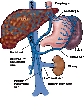
PANCE REVIEW: Varices

Let's head north of the heart for a while - I'm CV-system'd out for a bit. The esophagus. The must-know topics about the esophagus are below. I won't get to all of them on my blog... but you should def get to them in your studies!

Esophagitis
Motility d/o's
Mallory-Weiss tears
Neoplasms
Strictures
Varices

Let's chat about Varices today...

Def:
Dilations of veins (generally found distally)

Causes:
-Usual underlying cause is portal HTN which is usually secondary to cirrhosis
-Chronic viral HEP and NSAIDS can worsen bleeding
-*Budd-Chiari Syndrome may cause thrombosis of portal vein which can lead to varices

Dx:
-Usually diagnosed clinically
-Asymptomatic until they start to bleed - then they are LIFE THREATENING!

Tx:
-Hemodynamic support
-High vol IVF
-Vassopressors
-Endoscopic therapy+Pharm vasoconstriction

**30% of pts die during the 1st bleed, 50% of those that survive will die during the 2nd bleed**


Picture: http://www.bio.ri.ccf.org/Henderson/port.html
Source: AAPA/PAEA Exam Review Book

7.19.2012

PANCE REVIEW: Ischemic Heart Dz

Angina
1-Stable = < 3min during activity, better with rest

2-Unstable = > 30 min at rest

3-Prinzmetal = vasospasm at rest
Risk Factors (10):
-male
-increased age
-decreased estrogen state
-smoking
-fam hx
-HTN
-DM
-obesity
-dyslipidemia
-inactivity

Tests/Labs:
-EKG: horizontal or downslopping  ST seg (depression)
-Exercise Test: good non-invasive test
*Pimping Question: What signifies a positive exercise test? (Answer below)
-ECHO: prognostic indicator

Tx:
-sublingual nitro is the primary pharm tx
-chronic angina = beta blockers (prolong life)
-CCB decrease cardiac muscle O2 demand
-Platelet inhibition agent (aspirin, clopidogrel, ticlopidine)
-NOTE: Nitro and CCB only for Prinzmetal! Beta-blockers can provoke a spasm!


Answer: ST segment depression of 1mm


Source: AAPA and PAEA Exam Review Book

7.18.2012

PANCE REVIEW: Urgency vs Emergency

HTN, both primary and secondary, are fair game for the PANCE - but HTN in general is a huge topic. So in the interest of keeping these entries short and sweet, I went with the niche topic of urgency vs. emergency.

Hypertensive Urgency
Hypertensive Emergency
Systolic > 220, Diastolic >125
Diastolic > 130
Lower in HOURS
Lower within 1 HOUR
Complications: optic disc edema, end organ complications
Complications: hypertensive encephalopathy, IC hemorrhage, aortic dissection, pulm edema

Tests/Possible results:
1-EKG: heart failure/LVH
2-CXR: ventricular hypertrophy
3-Labs: decrease in Hbg/Hct, increase in BUN/Cr/Glucose - renal dz? DM? end organ damage?

Treatments:
Parenteral agents
-sodium nitroprusside
-if MI present, nitro or Beta-blocker
-if aortic dissection present, nitroprusside + beta blocker (Labetalol)


Source: AAPA and PAEA Exam Review Book

7.17.2012

PANCE REVIEW: Pancreas

Need to knows about the pancreas:

1. Acute pancreatitis
Notes:
Causes = ETOH abuse, cholelithiasis, hyperlipidemia, trauma, drugs, 2ndary to HIV meds
Clinical:
Classic presentation = epigastric pain radiates to back, N/V/F, pain alleviated by fetal position or leaning forward, leukocytosis, severe hypOvolemia
Labs/Tests:
-Increase serum amylase (not very helpful because it can be nl after 48-72hrs)
-Serum LIPASE = more sensitive/specific, but only with increases of 3x+
-Increase in liver enzymes if biliary obstruction
-Ranson's Criteria
Treatment:
-NPO
-IVF
-TPN
-pain management (MEPERIDINE (Demerol))
-consider antibx
-monitor for complications

2. Chronic pancreatitis
Notes:
-90% caused by ETOH abuse
- Triad only seen in 20% of pts [pancreatic calcification, steatorrhea, DM]

Clinical:
-same as Acute Pancreatitis +  steatorrhea (poor fat absorption)
Labs/Tests:
-serum lipase
-Abd film shows calcification in 20-30% of pts

Treatment: 
 -same as Acute Pancreatitis
-low fat diet at discharge
-NO ETOH

3. Pancreatic neoplasms
Notes:
-5th leading cause of CA related death in US
-Risks = incr age, obesity, tobacco, chronic pancreatitis, abd radiation, fam hx

Clinical:
-abdominal pain
-COURVIER'S SIGN (palpable gall bladder)

Labs/Tests:
-CT of abdomen
-angiography

Treatment: 
-surgical resection if no mets (Whipple procedure)
-POOR PROGNOSIS


Source: AAPA and PAEA Exam Review Book

7.16.2012

PANCE REVIEW: Congenital Heart Dz

According to the NCCPA site - the following congenital heart dz are fair game on the the PANCE. I have listed them with some key buzz words:

Red = non-cyanotic
Blue = cyanotic

Ventricular septal defect
-MOST COMMON
-systolic murmur, LLSB
-sx are size dependent (range from asymptomatic to CHF)
-"Outlet VSD" - most common in Chinese and Japanese

Atrial septal defect
-2nd most common
-systolic ejection murmur, 2nd LICS
-failure to thrive
-RV heave
-wide fixed S2 split 
 (If you can remember ASD is #2 - then you can remember 2nd LICS and fixed S2 split!)

*septal defects are #1 and #2 most common 

Coarctation of the aorta
-systolic, LUSB (may be Continuous)
-infants may present with CHF
-older kids may present with systolic HTN +/- murmur

Patent ductus arteriosus (PDA)
-continuous machinery murmur (I remember it has a PDA- like a phone- is a machine.)
-Wide pulse pressure (machine = "wide" screen TV)
-hyperdynamic apical pulse

Tetralogy of fallot
-crescendo-decrescendo
-HOLOsystolic at LSB 
-cyanosis, clubbing
-increased RV impulse at LLB
-Loud S2
-Associated things: polycythemia vera + hypercyanotic spells (Med Emergency!)


Source: AAPA and PAEA Exam Review Book
Image: http://www.healthofchildren.com/C/Congenital-Heart-Disease.html

7.12.2012

PANCE Review: Postual HypOtension (aka Orthostasis)

I have slowly begun to start studying for my boards... well at least organized my thoughts around studying for the PANCE... so many of my next posts will be dedicated to PANCE topics. I highly recommend visiting the official NCCPA site to get a list of the covered topics so help focus your studying. If it isn't on the Blueprint list, it isn't going to be on the exam. PANCE Blueprint site

Let's start simple:
Postual HypOtension (aka Orthostasis)

Def: A >20mmHg drop in SYSTOLIC BP between Supine and Sitting and/or Standing positions

If accompanied by an INCREASE in pulse by 15bpm = likely cause is DECREASE in BLOOD VOL (bleeding, dehydration, etc)

If is not accompanied by an increase in pulse = likely causes are meds or peripheral neuropathies

Tx: treatment is directed at the cause!


Source: AAPA/PAEA Exam Review Book

7.11.2012

ANKLE-BRACHIAL INDICES

ANKLE-BRACHIAL INDICES (ABI)
Indications: Presence of peripheral arterial disease
Technique
1. Have patient in supine position so arms, legs and heart are at same level.
2. Use blood pressure cuff and Doppler to measure systolic BP in both arms and record.
3. Use Doppler to identify location of dorsalis pedis and/or posterior tibialis pulses, mark location bilaterally.
4. Wrap BP cuff around lower leg. Using Doppler to listen to signal, inflate cuff until signal disappears, then slowly deflate until pulse signal returns. Record pressure at which pulse is heard (systolic) by Doppler at DP and PT in both ankles.
5. To calculate the AAI divide the highest SBP from each ankle (either DP or PT) by the highest SBP reading from the upper extremities.
6. ABI = ankle / arm systolic pressure
> 1.3 = suggests noncompressible, calcified vessels
0.91-1.3 = normal
0.41-0.9 = mild to mod peripheral arterial dz (range for claudication)
<0.4 = Severe peripheral arterial dz. (range for critical leg ischemia and rest pain)


Source: http://students.washington.edu/aomega/procedures.shtml#chestTube

7.05.2012

Chest Tubes

Today we put in a chest tube on a patient with about a 20-25% pneumo. Thought I'd share what I found in my research leading up to placing the tube:


Indications: Pneumothorax, hemothorax, empyema, recurrent pleural effusion
Contraindic.: Bleeding dyscrasia, anticoagulation, empyema caused by AFB
Technique
1. Obtain informed consent
2. Check coags / platelets
3. Consider sedating patient (painful)
4. Use 18-20 French tube for pneumothorax, 32-36 French tube for fluid or hemothorax
5. Assemble suction/drainage equipment and connect to suction
6. Position patient in supine position, elevate head of bed 30-60 degrees. Usual insertion site is at anterior axillary line at 4th or 5th intercostals space. Mark site.
7. Prep and drape in sterile fashion. Wear gown and mask.
8. Anesthesia at pleural insertion site: anesthetize skin over rib using 2 gauge needle, 10 cc syringe, 1% lidocaine. Anesthesia at incision site (rib below rib of pleural insertion). Using 22 gauge needle and 1% lidocaine, infiltrate subQ, muscle, periosteum, and parietal pleura.
9. Make 2-4 cm incision through skin and tissues over rib. Extend incision with blunt dissection using Kelly clamp, working towards superior aspect of rib above tunneling the course of the chest tube before entering the chest cavity.
10. Push Kelly clamp through parietal pleura. Inside pleural cavity, open clamp, then withdraw. Air or fluid should rush out.
11. Check to see that pleural space has been entered with finger.
12. Grasp chest tube with curved clamp. Clamp free end of chest tube with another clamp.
13. Place tube in pleural space. Direct tube superior, medial, posterior for fluid drainage. Direct tube superior and anterior for pneumothorax. All ventilation holes need to be in pleural space.
14. Attach end of tube to suction/drainage.
15. Use 1-0 or 2-0 silk or nylon to suture chest tube in place.
16. Cover site with 4x4 gauze (with Y cuts to fit around tube)
17. Tape gauze and tube in place
18. Obtain CXR to confirm placement
19. Remove chest tube when there is less than 150cc of fluid in 24hrs and no air leak.


Source: http://students.washington.edu/aomega/procedures.shtml#chestTube