Source: http://abnormalfacies.wordpress.com/2012/02/20/running-subcuticular-suture-technique/
Showing posts with label emergency medicine. Show all posts
Showing posts with label emergency medicine. Show all posts
4.23.2014
Subcuticular Suturing
I came across this blog post on how to do a subcuticular closure. It is well written with step by step pictures so why reinvent the wheel. I am just going to repost. Enjoy!
9.09.2013
Antibiotics for Appendicitis?
I have had quite the hiatus from blog entries recently. Life gets busy somehow. Ha. I recently had hip surgery and had some blog worthy experiences as a patient that I hope to write about soon - but for now, I came across this interesting article on a PA (Andrew Gray, PA-C) that refused an appendectomy in lieu of antibiotic treatment for his acute appendicitis. He made his choice based on the fact that he did not have insurance and the results of a Swedish study. It is a short read, but very interesting and have evoked some feisty comments.
Saving My Appendix: http://www.pulsemagazine.org
Saving My Appendix: http://www.pulsemagazine.org
7.14.2013
ACA Stroke Basics
Anterior Cerebral Artery (ACA) Stroke
The Anatomy


What deficits might you expect to see in a patient?
Source: http://www.neuroanatomy.ca/stroke_model/aca_info.html
Photo sources: http://missinglink.ucsf.edu/lm/ids_104_cns_injury/response%20_to_injury/watershed.htm
The Anatomy


What deficits might you expect to see in a patient?
- contralateral leg weakness (both motor and sensory), frontal lobe behavioral issues, +/- aphasia if prefrontal cortex involved, grasp reflex
Where does the ACA receive its blood supply from?
- Carotid arteries
Source: http://www.neuroanatomy.ca/stroke_model/aca_info.html
Photo sources: http://missinglink.ucsf.edu/lm/ids_104_cns_injury/response%20_to_injury/watershed.htm
7.08.2013
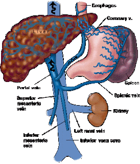
Causes of Renal Failure
Causes of Renal Failure broken down by pre-renal, renal, and post-renal.

Source: Clinical Survival Guide for PA Students by G.Broughton, MD, PhD
6.12.2013
What if PAs Couldn't Suture Anymore?
This a repost from a call sent out by the AAPA based on a resolution brought to the AMA's House of Delegates. Read it and take part in your future scope of practice as a PA.
While workforce experts are predicting a shortage of providers with the implementation of the Affordable Care Act, the American Medical Association Board of Trustess is proposing a resolution for its House of Delegates that could severely restrict PAs' ability to provide care to patients. PAs should be very concerned about the resolution, which will be considered by the AMA HOD when it convenes on Saturday, June 15. The recommendations are very restrictive and display a misunderstanding of the way PAs and doctors provide care as part of a team. Among other restrictions, the resolution expands the definition of surgery to include repair and removal of human tissue and, although parts of the resolution are somewhat unclear, states that surgery as defined in the resolution is to be performed ONLY BY PHYSICIANS. If adopted as presented, the resolution will call into question procedures like suturing, punch biopsies and vein harvesting, which PAs perform on a daily basis across many medical specialties. The resolution also proposes that only physicians should perform invasive procedures that utilize radiologic imaging. You can read Report 16 of the AMA Board of Trustees in full here.
Join AAPA in responding to negative AMA resolution
AAPA is spreading the word about the negative impact this resolution would have on patient care and PA practice, but we need your help. Please review the list of AMA delegates in your state or specialty, and if you have a connection, please let that physician know the true damage that this resolution could create. Also, talk with and encourage physicians in your practice to speak with other physician leaders about the resolution. AAPA's suggestion is that the resolution should be defeated, or modified to specifically state that it does not apply to PAs practicing within the parameters of state law.
For more information on the AMA resolution please contact Ann Davis, PA-C, MS, Senior Director of Constituent Organization Outreach and Advocacy, at ann@aapa.org.
5.20.2013
Atrial Fibrillation
AFib, The Basics
Characteristics
- irregularly iregular
- irregular RR intervals
- not a P wave in front of every QRS
- atrial rate = 400-600bpm, ventricular rate = 80-160bpm

P = pulmonary (COPD, PE)/pheo/pericarditis
I = ischemic heart dz +/- HTN
R = rheumatic heart dz
A= anemia/atrial myxoma
T = throtoxicosis
E = ethanol ("holiday heart)/cocaine
S = sepsis (post-operative)
Signs/Symptoms
- fatigue (most common)
- tachypnea
- palpitations
- lightheaded
Work Up
(Test yourself... why would you order each of these? what are you looking for?) - answers below
- EKG
- ECHO
- TSH (?)
- Baseline coags

- EKG = narrow complex QRS (<120msec), variable RR, irregular or absent P waves
- ECHO = maybe thrombi, maybe dilated L atrium
- TSH (?) = hyperthyroidism can cause AF
- Baseline coags = getting baseline prior to starting anticoagulation
Of note: if you are looking for THROMBI..."normal" ECHOs (transthoracic) has low sensitivity - transesophageal ECHOs allow for better visualization of L atrial appendage (location where most thrombi form)
source: First Aid for the Wards by Le, Bhushan, Skapik
pic source: http://www.saintvincenthealth.com/Services/Heart/Heart-Resource-Library/Atrial-Fibrillation/default.aspx
5.14.2013
Diabetes Insipidus, Part 2

Diagnosing DI
Polyuria = urine vol > 3L in 24 hrs - there are many causes of polyuria and it is important to figure out if the cause is DI or something else prior to establishing treatmentUrine osmolality (osm) of > 300 mOsmol/kg + high serum glucose --> think diabetes mellitus
Urine osmolality (osm) of > 300 mOsmol/kg + high serum urea --> think renal dz
Urine osmolality (osm) of < 200 mOsmol/kg + polyuria --> think DI
So you have a patient that has urine ohm < 200 + polyuria and you are thinking DI... how do you differentiate between central DI and nephrogenic DI?
Answer: water deprivation test
Findings:
Central DI
urine osm < plasma osm after dehydration
after ADH injections urine osm increases by >50%
Psychogenic DI
urine osm > plasma osm after dehydration
after ADH injections urine osm increases minimally
Nephrogenic DI
urine osm < plasma osm after dehydration
after ADH injections urine osm increases by <50%
Source:
Makaryus/Mcfarlane. DI: diagnosis and treatement of a complex disease Cleveland Clinic Journal of Medicine Jan 2006 Vol 73:1
pic source: medicaltextboks.blogspot.com
3.31.2013
PANCE Study Material: Male Reproductive Jargon
PANCE Study Material: Male Reproductive Jargon
HypOspadias = abnormal urethral opening underneath (remember hypO means "below")
Epispadias = abnormal urethral opening above (remember epi means "above")
*Phimosis = foreskin is too tight to retract over glans
*Paraphimosis = foreskin is too tight to return back to its usual position
*Usually congenital, but can be caused by trauma or infectious scarring
Source: Hardcore Pathology by Wahl
3.08.2013
Transcranial Doppler

- uses low-frequency U/S to evaluate flow velocity in cerebral vessels
Why might you order a transcranial doppler?
- evaluation of basal cerebral arteries
- finding spasms intercranially
- evaluating the patency of the MCA (you would look for this in patients with carotid stenosis)
What the upside to transcranial dopplers?
- no radiation
- fast
- non-invasive
- can evaluate the circle of willis/intercranial carotids
What is the downside?
- the outcomes is dependent on the person doing the test - skill and bias can play a role
Transcranial Doppler Video (a little old school - but good information)
Pic source: http://spencertechnologies.com
Source: Ferri's Best Test: A practical guide to clinical lab and medicine diagnostic imaging, Fred Ferri
3.06.2013
Hematuria Workup
A diagnostic algorithm to consider when working up hematuria...

Source: Ferri's Best Test: A practical guide to clinical lab and medicine diagnostic imaging, Fred Ferri

click on picture to enlarge
Source: Ferri's Best Test: A practical guide to clinical lab and medicine diagnostic imaging, Fred Ferri
3.03.2013
IV Therapy Complications

Thrombophlebitis - usually manifests with erythema, inflammation, and/or pain at the IV site (think about changing the IV q3 days to help prevent this)
Infiltration - this happens when whatever you are giving through the IV (meds/fluids) starts to leak into the surrounding tissue (this can cause a big problem - compartment syndrome - if the volume is large enough)
Blockage - something, a blood clot for example, can clog the IV making it unusable (flushes can help minimize the risk of this)
Air embolus
Source: Step up to Medicine 2nd ED Agabegi and Agabegi
Pic source: http://en.wikipedia.org/wiki/Intravenous_therapy
2.01.2013
Repleting K+

When thinking about repleting a potassium deficiency consider the following:
- Goal is of K is greater than 4 in any pt with active cardiac problems
- If your pt has nl renal function: 10mEq of KCl (IV or PO) will increase serum K by about 0.1mEq/L (so if your pt is at 3.6, about 40 mEq of KCl should help correct your patient)
- Don't replete if patient is on dialysis (consult the dialysis team)
- f your pt has compromised renal function: divide the mEq of normal repletion by the pt's Cr (example: pt has Cr of 3, then you use 1/3 of the nl repletion amount - so instead of 9mEq, you'd use 3mEq)
- PO can cause nausea
- IV can be painful
- Typical combos: 10mEq/100cc or 10mEq/50cc (peripheral IV) and 20mEq/50cc (central line)
- Be careful when repleting its with renal insufficiency or in pts with high risk of tumor lysis
*This is not meant to substitute for clinical judgement, just suggestions to think about when treating K+ deficiencies in your patients.
Source:
http://www.eric.vcu.edu/home/curriculum/print/Intern_Ward_Survival_Guide_2009.pdf
http://www.surgicalcriticalcare.net/Guidelines/electrolyte_replacement.pdf
photo: http://mattrosenart.deviantart.com/art/Potassium-195578504
1.08.2013
Brown-Sequard syndrome
Brown-Sequard... Jekyll-Hyde.... that's kind of how I remember what it is. Two different sides to the spinal cord, one good (uninjured) and one bad (injured). Brown-Sequard syndrome is characteristically seen in penetrating injuries (but can be seen in unilateral blunt injuries as well). The syndrome results in one side of the spinal cord being injured.
What do you see clinically?
Motor, position, and vibration are gone on the SAME side as the injured spinal cord (these tracts cross at the brainstem)
Pain and temperature are gone on the OPPOSITE side of the injured spinal cord (these tracts cross at the cord at or near the level of innervation)
What to do clinically?
The diagnosis is generally made clinically based on hx and physical. This warrants a neurosurgical or orthopedic consult and MRI if requested. Lastly, studies have shown that high doses of steriods (methylprednisolone) decreases inflammation by suppressing polymorphonuclear leukocytes and reversing increased capillary permeability.
Sources:
1.http://emedicine.medscape.com/article/791539-workup#a0720
2.Abernathy's Surgical Secrets by Harken and Moore
What do you see clinically?
Motor, position, and vibration are gone on the SAME side as the injured spinal cord (these tracts cross at the brainstem)
Pain and temperature are gone on the OPPOSITE side of the injured spinal cord (these tracts cross at the cord at or near the level of innervation)
 |
| http://nhananhana.wordpress.com/2010/01/28/brown-sequard-syndrome/ |
The diagnosis is generally made clinically based on hx and physical. This warrants a neurosurgical or orthopedic consult and MRI if requested. Lastly, studies have shown that high doses of steriods (methylprednisolone) decreases inflammation by suppressing polymorphonuclear leukocytes and reversing increased capillary permeability.
Sources:
1.http://emedicine.medscape.com/article/791539-workup#a0720
2.Abernathy's Surgical Secrets by Harken and Moore
11.19.2012
Free Book for Your Surgical Rotation
Scribd is a great resource for free textbooks (and not just the cheap ones). You can buy a subscription to have total access (Premium) or you can upload non-copyrighted materials and get a Premium membership for free.
I found this surgical book written for students on their surgical rotations. It is pretty awesome and comprehensive. A snippet from the table of contents is below.

I found this surgical book written for students on their surgical rotations. It is pretty awesome and comprehensive. A snippet from the table of contents is below.

8.12.2012
Asthma Charts
Every PACK-RAT I've taken has had at least 1-2 questions on asthma. During my primary care, pediatrics, and emergency medicine rotations I keep these charts with me because I used them daily.


Source: http://www.rtmagazine.com/issues/articles/2009-05_01.asp, http://www.uspharmacist.com/content/c/10133/?t=men%27s_health,otc_medications


Source: http://www.rtmagazine.com/issues/articles/2009-05_01.asp, http://www.uspharmacist.com/content/c/10133/?t=men%27s_health,otc_medications
8.08.2012
Reading a Chest Xray
You should feel confident reading a chest x-ray (CXR). It is one of the few films that will follow you from rotation to rotation. It doesn't matter if it is pediatrics, internal medicine, or surgery - You need to know how to read a CXR. Below are a couple sources to choose from because not everyone teaches or learns this in the same way. Here are a couple tips that I learned during my rotations from studying, my preceptors, or just plain screwing up!
- The first thing you should check is the name/date/type of film! (On one of my rotations, an intern (1st yr resident) was asked to read a chest X-ray for one of our patients who had just gotten a chest tube placed. He did a great job with lung pathology and describing the fluid - and he was also able to pick out that the chest tube was perfectly placed. I was impressed until the chief resident said "great job, you just harmed your patient." The chief had purposefully put up a CXR from 2 years ago when the pt had rec'd another chest tube. He then pulled up the current CXR to reveal that the tube was improperly placed. )
- Read every film in the same order every time.
- Learn the anatomy of what you are reading.
University of Washington's Method
1. PA or AP, supine or upright
2. Pt rotated? Check for vertebral and clavicle symmetry.
3. Lung volumes
4. Tube & line placement
- ETT 3-5 cm above carina
- NGT in stomach
- FT in stomach/duodenum
- Central line in SVC/R atrium
- Swan in PA
5. Pneumothorax: check apices on upright film, deep sulcus sign at bases
6. Pleural effusion, pleural thickening
7. Mediastinum: normal contour, wide
8. Heart: normal size, cardiomegaly
9. Lung parenchyma: masses, opacites, look for silhouette sign
10. Soft tissues: foreign bodies, SQ air, breast shadows
11. Bones: fractures, osteopenia, abnormalities
Silhouette Sign = obscuring of normal borders on radiograph caused by intrathoracic lesion.
Obscured R heart border = R middle lobe
Obscured L heart border = Lingula
Obscured diaphragm = Lower lobe
8.06.2012
Lumbar Punctures
LUMBAR PUNCTURES
Indications: Suspected CNS infection, SAH, Guillain-Barre syndrome, MS, SLE
Measure intracranial pressure (pseudotumor cerebri)
Contraindications: Increased intracranial pressure (except to dx pseudotumor cerebri), supratentorial mass lesion, thrombocytopenia, bleeding dyscrasia
Complications: Post-LP headache, brain herniation if mass lesion present or increased intracranial pressure, bloody tap if venous plexus punctured.
Technique
1. Obtain informed consent
2. Position patient with back near edge of bed in lateral recumbent position. Have patient flex hips and draw knees up to chest to increase curvature of spine.
3. Palpate iliac crests and identify L3 and L4 interspaces.
4. Open tray, wear sterile gloves, and set up tubes in order, 1-4.
5. Prep and drape skin in sterile fashion
6. Infiltrate skin with 1% lidocaine
7. Use 20-22 gauge spinal needle. Insert at interspace with needle angled slightly toward umbilicus (cephalad). Keep level of needle in line with horizontal plane.
8. A course resistance can be felt as the needle passes through the paraspinous ligaments and a “pop” may be felt when needle passes through the dura.
9. Withdraw stylus fully to check for fluid.
10. Once fluid is obtained, place stopcock and manometer on hub of needle to obtain opening pressure.
11. Fill tubes in order, 2-3cc per tube
12. Once fluid has been collected, replace stylus and withdraw needle.
13. Cover site with sterile dressing and have patient remain lying down in supine position for 2 hours.
14. Observe tubes for occult blood. Decreasing amounts of blood in tubes 1-4 suggests a bloody tap, while increasing or steady amounts suggests an CNS bleed.
15. Send fluid for analysis:
Tube #1: glucose, protein, protein electrophoresis
Tube #2: Gram stain, culture, bacteria, fungal, TB, viral
Tube #3: cell count, differential
Tube #4: VDRL, India ink, cytology
7.20.2012
PANCE REVIEW: Varices
Let's head north of the heart for a while - I'm CV-system'd out for a bit. The esophagus. The must-know topics about the esophagus are below. I won't get to all of them on my blog... but you should def get to them in your studies!
Esophagitis
Motility d/o's
Mallory-Weiss tears
Neoplasms
Strictures
Varices
Let's chat about Varices today...

Def:
Dilations of veins (generally found distally)
Causes:
-Usual underlying cause is portal HTN which is usually secondary to cirrhosis
-Chronic viral HEP and NSAIDS can worsen bleeding
-*Budd-Chiari Syndrome may cause thrombosis of portal vein which can lead to varices
Dx:
-Usually diagnosed clinically
-Asymptomatic until they start to bleed - then they are LIFE THREATENING!
Tx:
-Hemodynamic support
-High vol IVF
-Vassopressors
-Endoscopic therapy+Pharm vasoconstriction
**30% of pts die during the 1st bleed, 50% of those that survive will die during the 2nd bleed**
Picture: http://www.bio.ri.ccf.org/Henderson/port.html
Source: AAPA/PAEA Exam Review Book
7.19.2012
PANCE REVIEW: Ischemic Heart Dz

Angina
1-Stable = < 3min during activity, better with rest2-Unstable = > 30 min at rest
3-Prinzmetal = vasospasm at rest
Risk Factors (10):
-male-increased age
-decreased estrogen state
-smoking
-fam hx
-HTN
-DM
-obesity
-dyslipidemia
-inactivity
Tests/Labs:
-EKG: horizontal or downslopping ST seg (depression)-Exercise Test: good non-invasive test
*Pimping Question: What signifies a positive exercise test? (Answer below)
-ECHO: prognostic indicator
Tx:
-sublingual nitro is the primary pharm tx-chronic angina = beta blockers (prolong life)
-CCB decrease cardiac muscle O2 demand
-Platelet inhibition agent (aspirin, clopidogrel, ticlopidine)
-NOTE: Nitro and CCB only for Prinzmetal! Beta-blockers can provoke a spasm!
Answer: ST segment depression of 1mm
Source: AAPA and PAEA Exam Review Book
7.18.2012
PANCE REVIEW: Urgency vs Emergency
HTN, both primary and secondary, are fair game for the PANCE - but HTN in general is a huge topic. So in the interest of keeping these entries short and sweet, I went with the niche topic of urgency vs. emergency.
Tests/Possible results:
1-EKG: heart failure/LVH
2-CXR: ventricular hypertrophy
3-Labs: decrease in Hbg/Hct, increase in BUN/Cr/Glucose - renal dz? DM? end organ damage?
Treatments:
Parenteral agents
-sodium nitroprusside
-if MI present, nitro or Beta-blocker
-if aortic dissection present, nitroprusside + beta blocker (Labetalol)
Source: AAPA and PAEA Exam Review Book
Hypertensive Urgency
|
Hypertensive Emergency
|
Systolic > 220, Diastolic >125
|
Diastolic > 130
|
Lower in HOURS
|
Lower within 1 HOUR
|
Complications: optic disc edema, end organ complications
|
Complications: hypertensive encephalopathy, IC hemorrhage, aortic
dissection, pulm edema
|
Tests/Possible results:
1-EKG: heart failure/LVH
2-CXR: ventricular hypertrophy
3-Labs: decrease in Hbg/Hct, increase in BUN/Cr/Glucose - renal dz? DM? end organ damage?
Treatments:
Parenteral agents
-sodium nitroprusside
-if MI present, nitro or Beta-blocker
-if aortic dissection present, nitroprusside + beta blocker (Labetalol)
Source: AAPA and PAEA Exam Review Book
Subscribe to:
Posts (Atom)